August's Case of the Month
Use of ultrasound to identify cause of canine hyperthyroidism found incidentally
Dr. Emily Evans
PATIENT INFORMATION:
Age: 12 years, 8 Months
Gender: Neutered Male
Species: Canine
Breed: Maltese Mix
Weight: 14lbs
HISTORY:
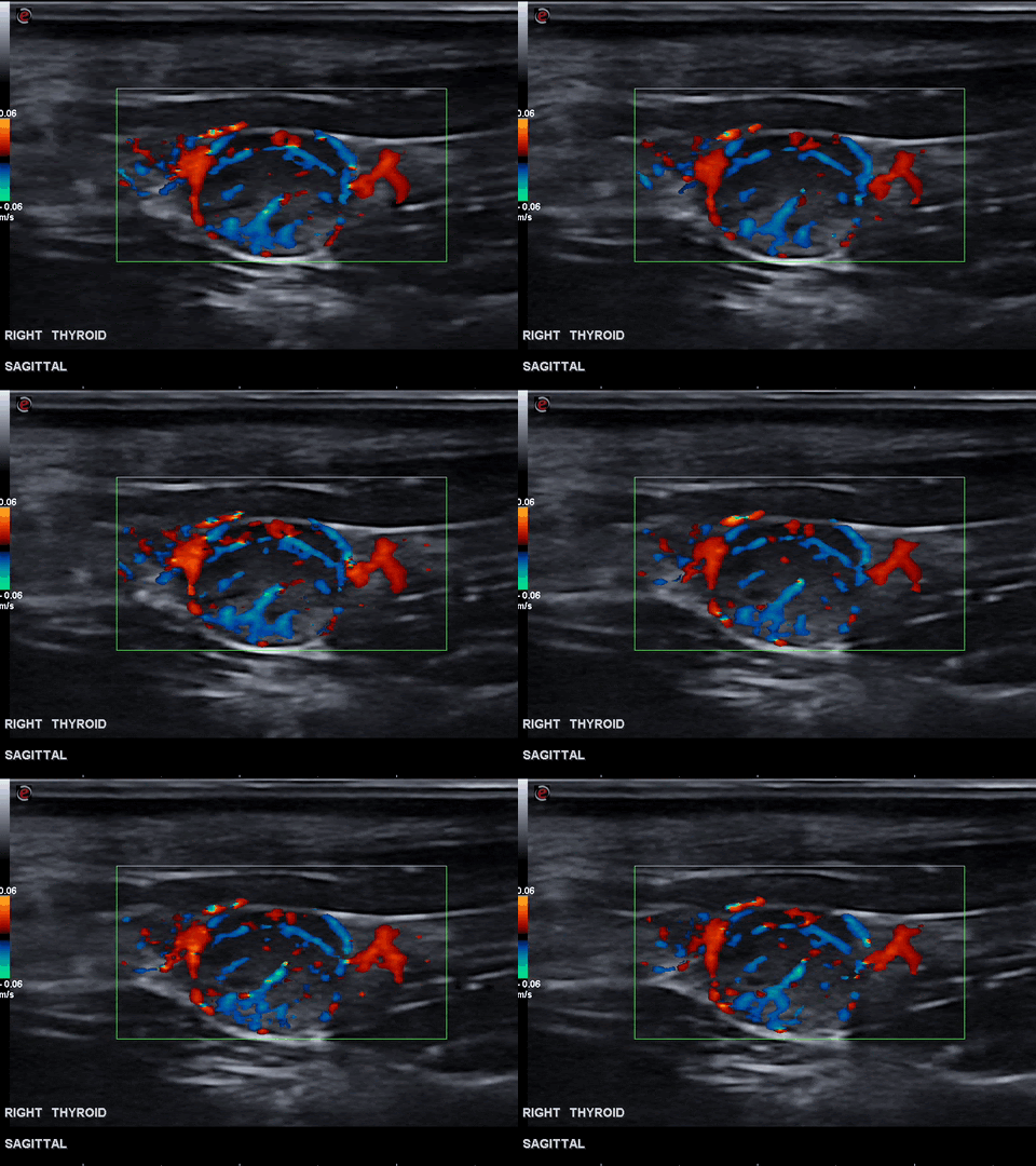
Patient presented(approximately 2-3 weeks prior to ultrasound) for a large draining tract/abscess on ventral neck. Antibiotic and NSAID therapy resolved the swelling and the patient returned to normal activity and behavior at home. Following resolution of abscess routine senior screening lab work revealed elevation in total T4 (4.0ug/dl, normal 0.8-3.5ug/dl). No masses or nodules were palpable on exam and the patient was found to be asymptomatic. Cervical ultrasound was requested. Findings on Cervical Ultrasound - the right thyroid gland had a hypoechoic nodule measuring 8.9 x 8.0mm that was highly vascular on doppler evaluation.
Image 1: Right thyroid and thyroid nodule measuring 8.9x8.0mm.
Image 2: Right thyroid nodule was found to be highly vascular on doppler evaluation. Thyroid tumors tend to be highly vascular.
Other findings of cervical ultrasound:
-- The left thyroid was found to be reduced in size. (Atrophied)
-- Submandibular lymph nodes were mildly enlarged and hypoechoic. Lt/Rt: 9.9x3.8mm/ 9.8x4.0mm
SONOGRAPHIC ANALYSIS:
Right thyroid nodule - the findings are moderate -DDx: thyroid carcinoma vs. benign growth vs. parathyroid tumor vs. other neoplasia. Note: It is not possible to differentiate parathyroid nodules from thyroid nodules without histopathology.
Lymph node changes - the findings are mild -DDx: reactive secondary to recent abscess vs. metastatic infiltration.
ADDITIONAL DIAGNOSTICS:
Fine needle biopsies of the thyroid nodule were collected under sedation on the day of ultrasound and submitted to Eastern VetPath.
Image 3: Clusters of cells with poorly defined cell borders are shown. Note: Thyroid tumor cytology is often hemodiluted due to vascular nature. (Image provided by Dr. Scruggs at Eastern VetPath.)
DIAGNOSIS:
Cytology results confirm diagnosis of a tumor of endocrine/neuroendocrine origin with 100% confidence. Given the anatomic location, elevated T4 and species of patient, there is a high clinical suspicion for a thyroid carcinoma.
Recent abscessation of the ventral neck was likely coincidental.
CASE OUTCOME:
At the time of this case report, the owner was still deciding the next steps to pursue.
Referral to a veterinary oncologist was recommended to provide the best therapeutic plan. Additional diagnostics should include surgical biopsy and histopathology, abdominal ultrasound, thoracic radiographs, and lymph node biopsy.
Since this mass appears to be identified early and is still small(under 1.0cm) we are hopeful that this patient will have a good outcome with appropriate therapy.
PROGNOSIS/DISCUSSION:
The thyroid nodule is suspected to be the cause of hyperthyroidism in this patient. Hyperthyroidism secondary to thyroid masses is a rare condition in dogs. Studies have indicated that most thyroid neoplasias are euthyroid(60%), 30% are hypothyroid (due to the destruction of normal thyroid tissue), and only 10% are hyperthyroid.1,2. Most thyroid tumors in dogs are large, malignant and non-functional.
Sonographer: Emily Evans, DVM
______
References -
1) Wucherer K L, Wilke V: Thyroid cancer in dogs: an update based on 638 cases (1995-2005). J Am Anim Hosp Asso 2010 Vol 46 (4) pp. 249-54.
2) Lunn KF, Page RL: Tumors of the Endocrine System. Withrow & MacEwen’s Small Animal Clinical Oncology, 5th ed. St. Louis, Saunders Elsevier 201 pp. 513-515.
Special thanks to our friends at VCA Pet’s First in Richmond, Virginia and Dr. Jennifer Scruggs PHD, DVM, DIPLOMATE, ACVP (CLINICAL & ANATOMIC PATHOLOGY) at EasternVetPath for this help with this case.


