January's Case of the Month
Sonographic Findings of Cor Triatriatum Dexter (CTD) in a Canine Puppy.
Dr. Shadawn Salmond-Jimenez
Patient Information:
Age: 2 months
Gender: Female Intact
Species: Canine
Breed: Labrador Retriever
History
Patient presented from another hospital where hepatomegaly was diagnosed on exam. A Grade II/VI murmur was ausculted on presentation. Patient was eating and drinking well with no clinical signs. Radiographs performed showed abdominal effusion (more than expected for the age). Thoracic radiographs showed dilation of the caudal vena cava, but otherwise normal thorax. An echocardiogram was requested and performed.
Echocardiogram Findings
A circular cystic structure is identified in the region of the left/right atrium. There is a trace of flow across the interatrial septum that is seen from several angles which likely represents flow through a patent foramen ovale.
Interpretation
The cystic structure is likely a cor triatriatum dexter (CTD) which is a congenital defect where the right atrium is divided into a lower pressure cranial chamber that communicates with the tricuspid valve and higher pressure caudal chamber which may be the cause of the right to left flow through the PFO (Persistent Foramen Ovale).
Monitoring and Therapeutic Recommendations
The CTD may be an incidental finding but if signs of right-sided failure develop – ascites, treatment may be needed. Treatment involves balloon dilatation of the membrane and in rare cases stent implantation. Follow up is recommended.
Case Outcome
Surgical options pending clinical symptoms have been discussed. Patient remains asymptomatic.
Brief Summary of Cor Triatriatum Dexter
Cor Triatriatum Dexter (CTD) has been infrequently reported in dogs and the overall prevalence is low [1]. With this cardiac anomaly, the embryonic right valve of the sinus venosus fails to regress, resulting in partitioning of the right atrium (RA) into two distinct chambers and effectively creating a triatrial heart [2].
Given the varying extent of regression failure seen in different cases, the venous return from the abdomen to the RA can be impeded to varying degrees. This may result in clinical signs suggestive of caudal right-sided congestive heart failure (CHF) or a Budd-Chiari-like syndrome [3].
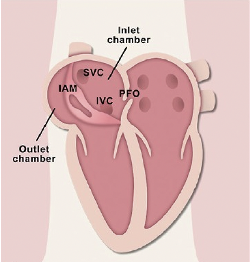
CTD can present with a variety of concurrent cardiac anomalies including persistent foramen ovale (PFO) (a hole between the left and right atria (upper chambers) of the heart).
For a positive long-term outcome balloon dilatation [5, 8, 19, 20], including cutting balloon techniques [20] or surgical correction under inflow occlusion or extracorporal circulation [2, 4, 10, 21, 22] have been reported as the treatment of choice.
https://www.ncbi.nlm.nih.gov/pmc/articles/PMC5210289/
Cor triatriatum dextrum (Human Heart Illustration). IAM: Intra-atrial membrane partitioning the inlet and outlet chamber (Right Atrium), IVC: Inferior vena cava orifice, PFO: Patent foramen ovale, PV: Pulmonary vein orifice, SVC: Superior vena cava (Annals of Cardiac Anesthesia)
Above: Echocardiogram demonstrating a right long axis view of patient. A Large rounded thick walled structure (cystic structure) (blue arrow and blue asterisk) at the cavoatrial junction is seen between both normal atria (right atrium shown by yellow arrow and left atrium shown by pink arrow)
Below: Normal echocardiogram demonstrating a long axis view with normal left atrium (pink arrow) and right atrium (yellow arrow)
Above: Apical four-chamber view of the patient’s heart showing the cystic structure characteristic of cor triatriatum dexter (blue arrow) in the region of the right atrium (yellow arrow) and left atrium (pink arrow).
Below: Apical four-chamber view showing normal right atrium (yellow arrow) and left atrium (pink arrow).
A special thanks to DVM STAT Consulting for the echocardiogram interpretation and consultation and the staff at Prince Georges Animal Hospital for this interesting case.


