June's Case of the Month - 2023
Confirmation of Rodenticide Toxicity
PATIENT INFORMATION:
Age: 7.5 years
Gender: Male
Species: Canine
Breed: Pitbull
Weight: 77lbs
HISTORY:
The patient presented for lethargy. There was an imprecise history given which included possible ingestion of multiple toxins from a garden shed (Potentially ingested agents - Diphacinone, bromethalin, pyrethroids, weed killer, possible other “garage” contents.) The exact timing of ingestion provided by the owner was unclear.
On initial physical examination the patient was dull and had a large caudal abdominal mass/bladder. There was concern for a bleeding tumor or other caudal abdominal mass on physical exam and radiographs.
ADDITIONAL DIAGNOSTICS:
Radiographs - Decreased serosal detail, mass effect in the caudal abdomen.
PCV/TS -initial presentation 36%/6.8 repeat evaluation 23%/4.8
ULTRASOUND FINDINGS AND IMAGES:
Spleen - Normal size(2.0cm), shape, and echogenicity. There is a heterogenous well defined capsule altering hypoechoic nodule in the spleen(2.0x2.6cm) The splenic nodule is vascular.
Liver -Normal size, shape and moderately coarse echogenicity. No focal lesions are appreciated. The gall bladder is moderately distended with normal anechoic and hyperechoic unorganized dependent bile that is not resulting in obstruction. No common bile duct dilation is seen.
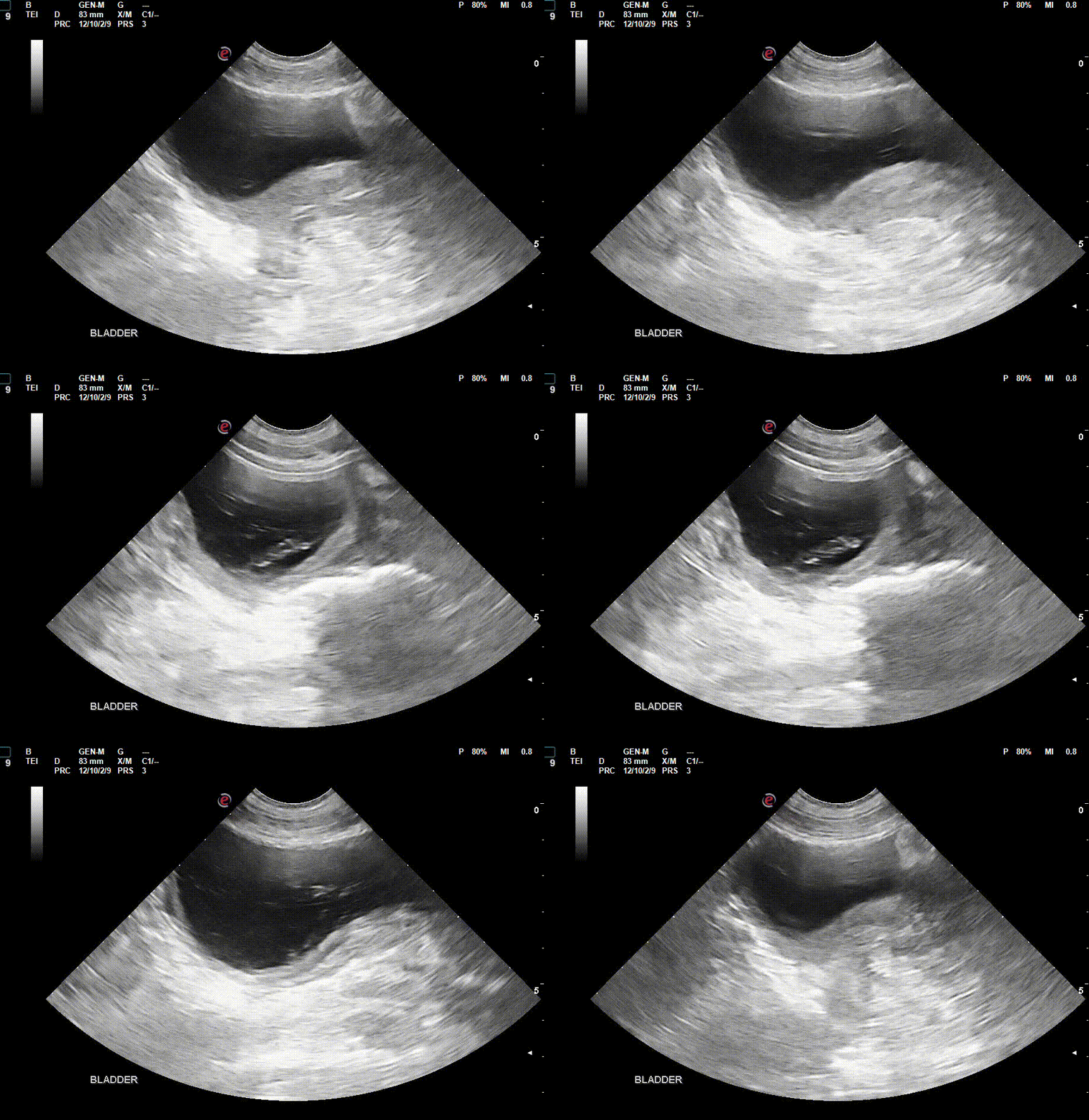
Serosal Surfaces- There is a mild-moderate amount of free fluid in the abdomen. Fat and tissue surrounding the prostate is severely heterogenous and hyperechoic. Mesentery throughout the abdomen is diffusely hyperechoic.
Kidneys- Normal size (Lt/Rt = 6.9/7.4 cm) and shape with normal corticomedullary dimensions. No pyelectasia visualized. There is a moderate amount of hypoechoic heterogenous avascular change in the retroperitoneal space bilaterally with a mild amount of free fluid.
Prostate- Severe generalized enlargement with hyperechoic coarse echogenicity and multifocal anechoic small cysts throughout. No mineralization or focal lesions. (LxDxW - 9.7x5.0x5.4cm)
Lymph Nodes - The medial iliac and sublumbar lymph nodes are mildly enlarged with rounded shape having homogenous mildly hypoechoic echogenicity. (Lt/Rt: 1.2/1.7cm, 0.7cm)
Urinary Bladder- The bladder is mildly distended with mildly echogenic urine and the bladder walls are mild-moderately thickened(10.2mm) and irregular in contour and thickness. No overt obstruction, uroliths, or neoplasia noted.
DIAGNOSIS:
Multiple sites of suspected hemorrhage and hematomas(ascites, retroperitoneal change, splenic lesions, urinary bladder and mesenteric changes) secondary to anticoagulant rodenticide ingestion.
Prostatic enlargement coupled with ascites and mesenteric change gave the appearance of a caudal abdominal mass. The prostatic change is likely secondary to prostatitis or benign prostatic hyperplasia in this intact individual.
CASE OUTCOME:
No samples were collected from this patient due to concerns for bleeding diathesis. The patient was transferred to ER for a transfusion and vitamin K therapy was initiated.
Follow up with the owners 5 days after the initial presentation, the patient was said to be doing well.
PROGNOSIS/DISCUSSION:
Recheck ultrasound 30 days after finishing was recommended to reassess changes.
Neuter was recommended to address prostatic enlargement.
DISCUSSION:
Abdominal ultrasound can be utilized in cases of suspected or known anticoagulant rodenticide toxicity to assess for presence of and extent of internal hemorrhage. This patient had no external signs of a bleeding diathesis but multiple intra abdominal signs which were concerning for hemorrhage and supported the diagnosis of anticoagulant rodenticide ingestion. This evidence aided in the initiation of appropriate therapy.
References:
Merola, V. (2002). Anticoagulant rodenticides: Deadly for pests, dangerous for pets. Veterinary Medicine.
Baranidharan GR, AP Nambi, S Kavitha, PS.Thirunavukkarasu , P Sridhar, H Yamini and S Muthuvel, 2014. Anticoa- gulant rodenticide toxicity and secondary haemostatic disorder in a dog. Inter J Vet Sci, 3(1): 37-39. https://url.avanan.click/v2/___www.ijvets.com___.YXAzOnN2cDphOmc6N2NiY2E1NzQ2MWJhNTFjZjRiNDgxNjIyZmEyY2E4MWY6NjpjZWJmOjM2NmE0M2FhOWViOTcxZjg5MDZkYWE2NGVjOTJmYWM3NTFjMmE1N2UwNTk4NDUxODgxY2Y3ZWRmNWIyMGNhYzY6dDpU
Sheafor, S.E.; Couto, C.G.: Anticoagulant rodenticide toxicity in 21 dogs. JAAHA 35 (1):38-46; 1999
RT O’Brien, E F WoodVet Radiol Ultrasound Jul-Aug 1998;39(4):354-6 Urinary bladder mural hemorrhage associated with systemic bleeding disorders in three dogs
Thank you to the veterinarians and staff of Varina Veterinary Clinic for collaborating with us on this case.


